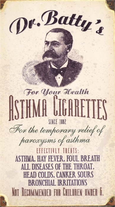
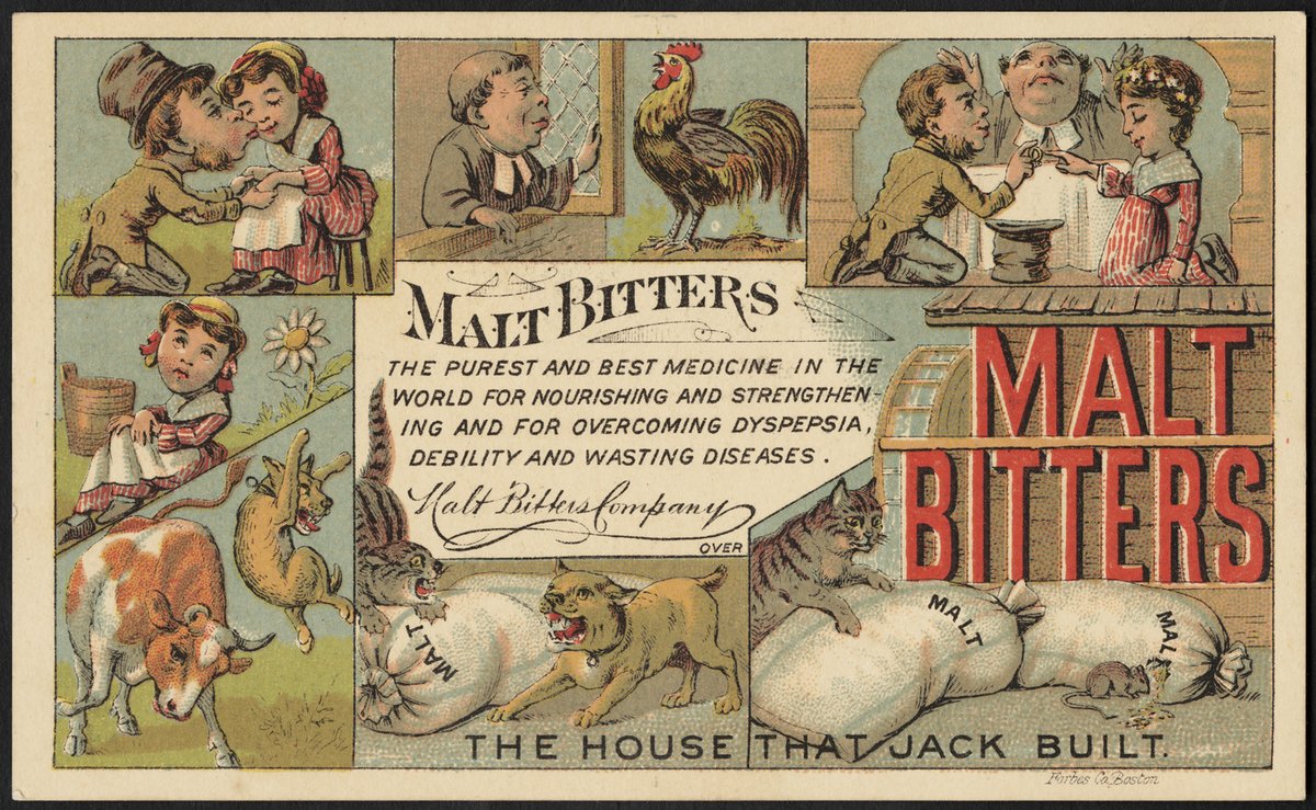
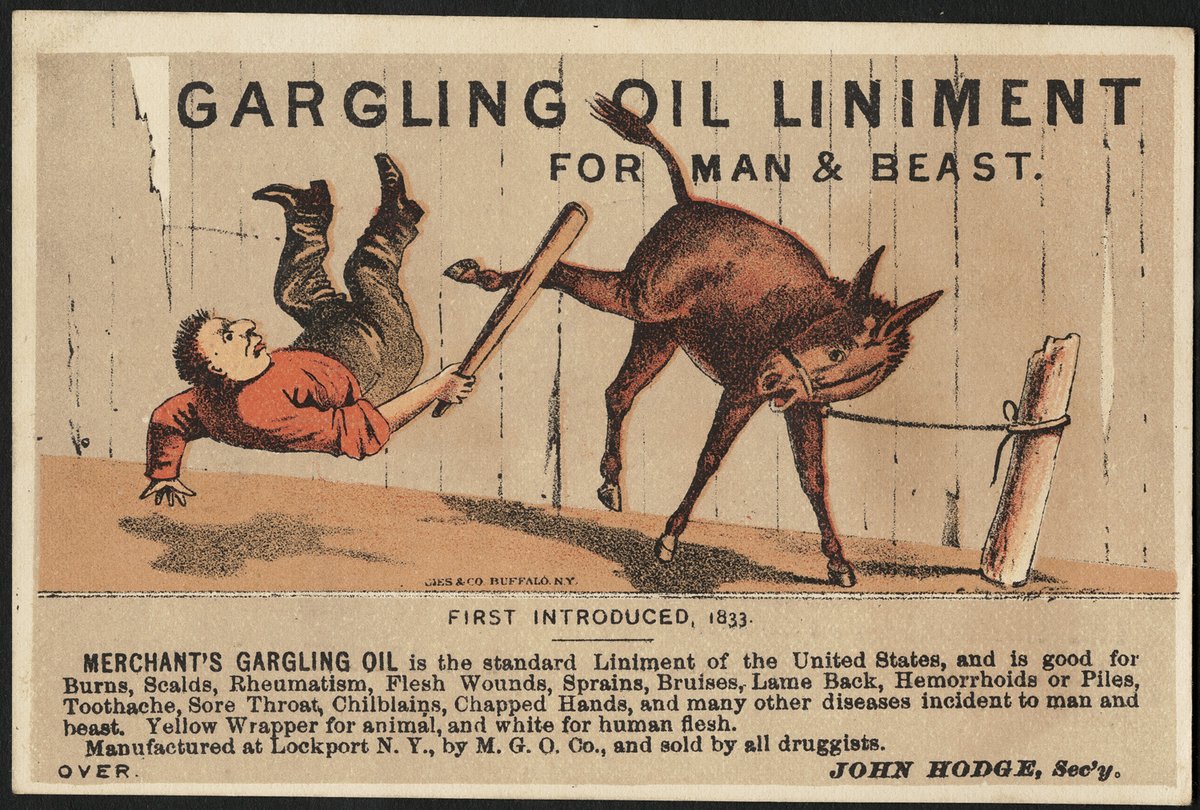
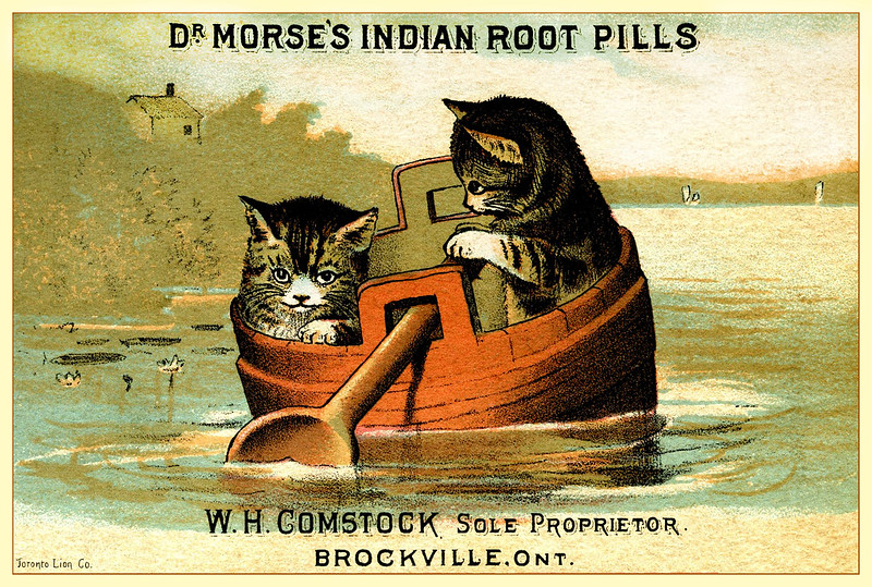
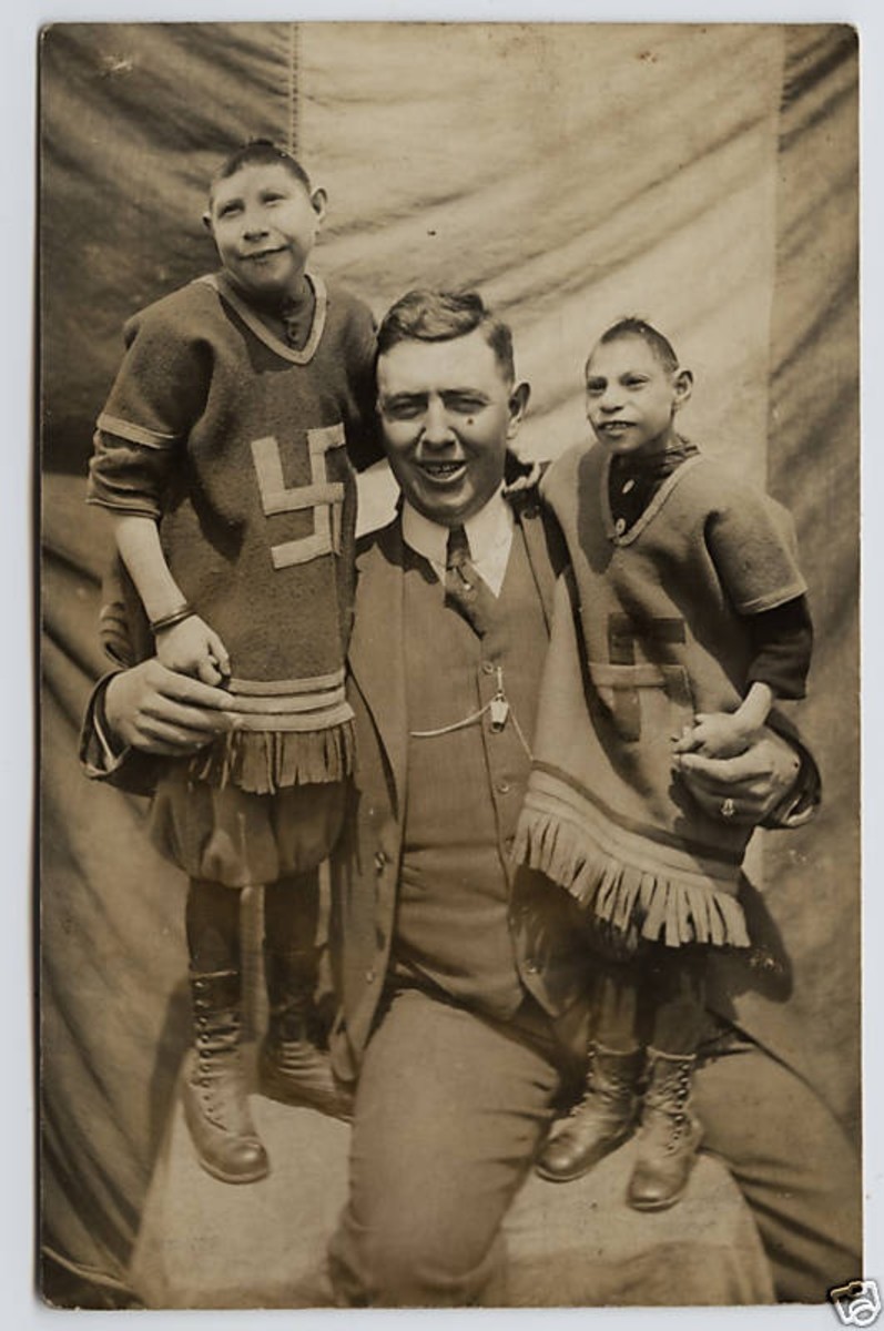
[Thread]: Old school "medicine" ads obviously created by aliens.
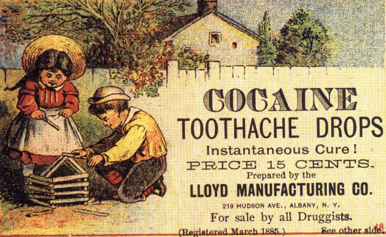
Let's begin with cocaine toothache drops, for only 15 cents.
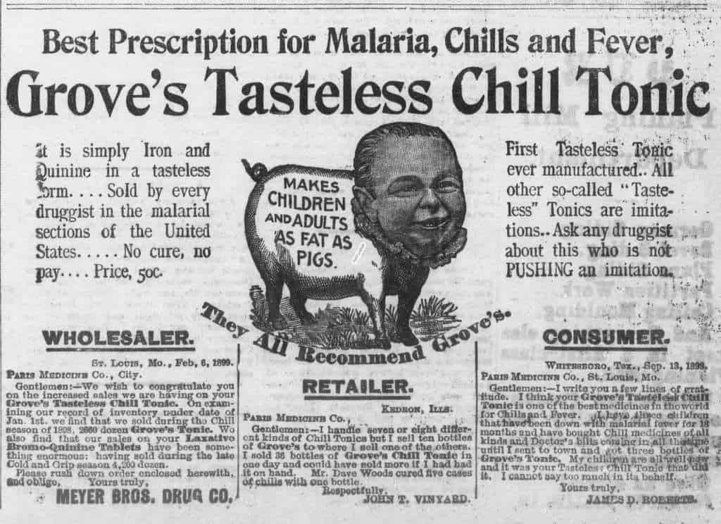
"It is simply iron and Quinine in a tasteless form".



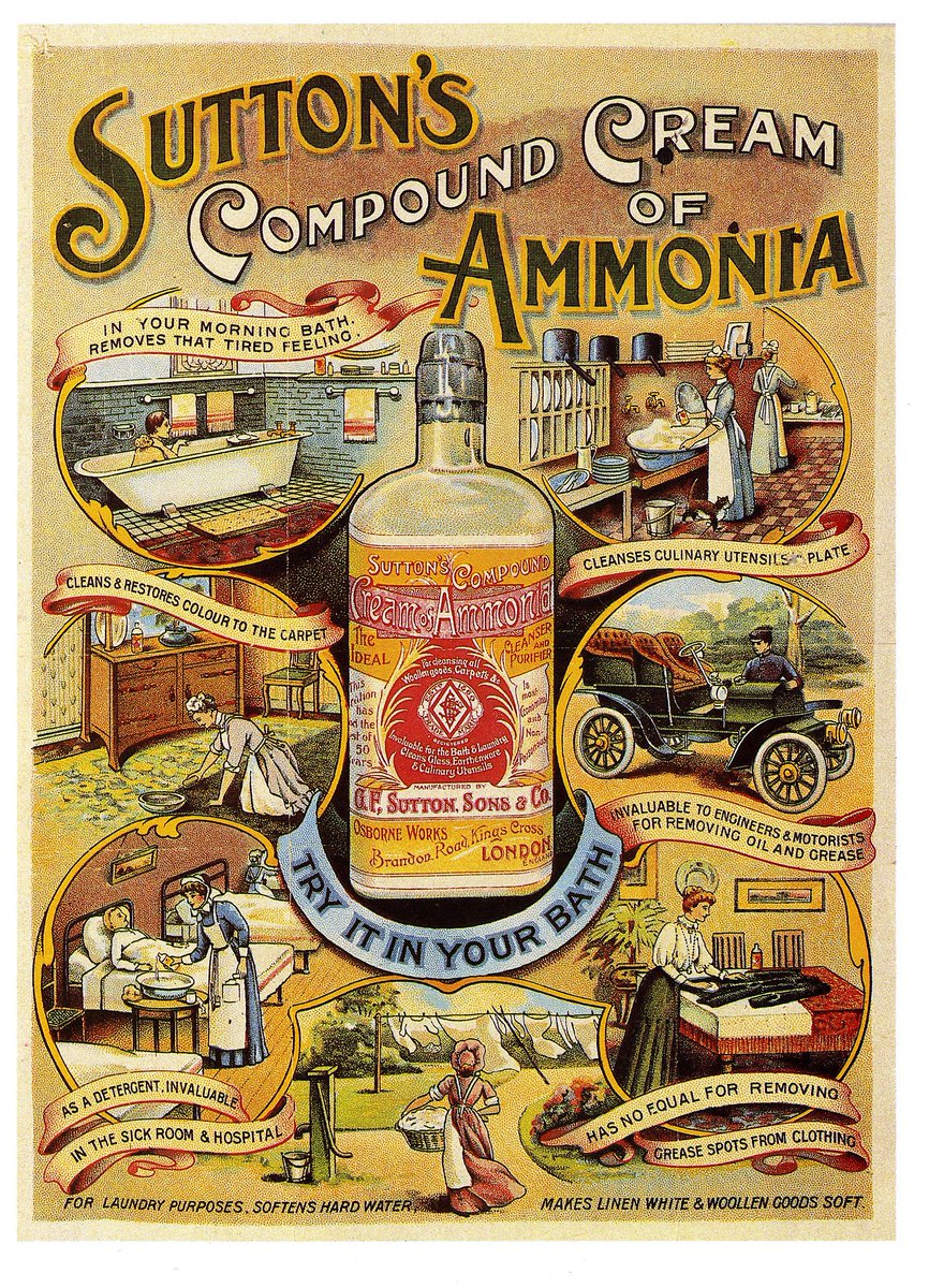
Don't forget to use it when you need to clean and restore color to the carpet.

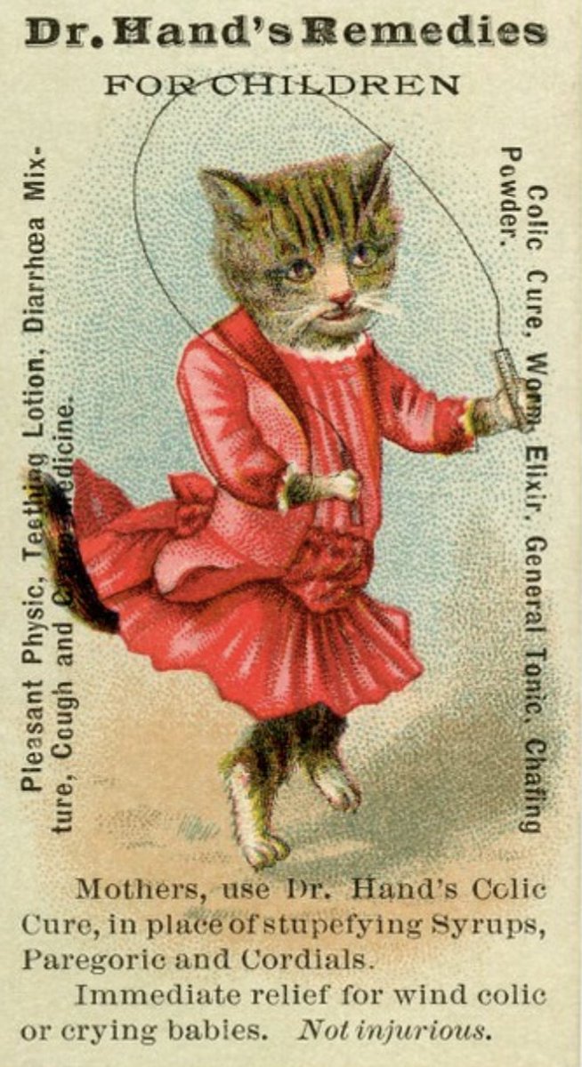
The cat in a dress skipping rope is important.


More from Marina Amaral








More from Health







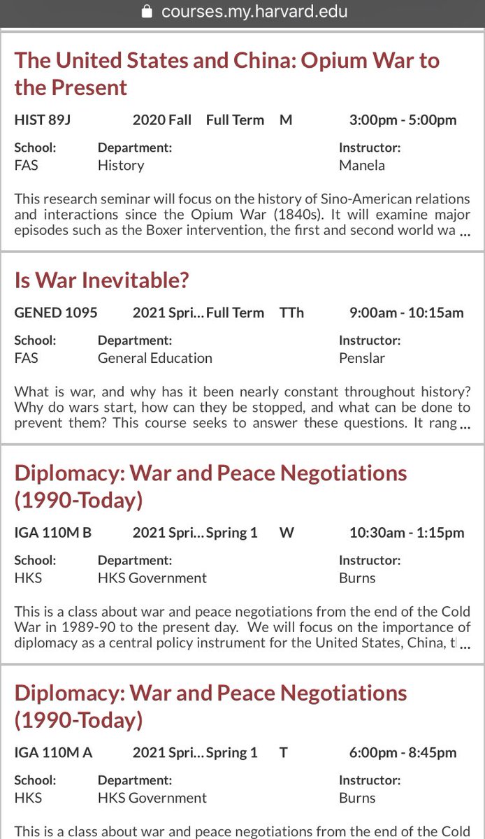
Now you know I love to sh-t in Harvard. But I also like accuracy. So I decided to go look at Harvard’s catalog to see its lack of military history that this article describes (they only teach history of pets it claims) and what I found shocked me! Shocked me! A thread: 1/
First off, Harvard students literally have multiple sections of military history that they can take listed. (It appears these ones are taught at MIT, so they might have to walk down the street for these) but... 2/

Say they want to stay on campus...they can only take numerous classes on war and diplomacy...3/
They have an entire class on Yalta. That’s right. An entire class on Yalta. 4/

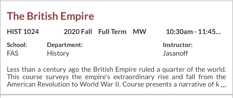
But wait! There is more! They can take the British Empire, The Fall of the Roman Empire for those wanting traditional topics... 5/
\u201cMilitary history\u201d is only in decline if you\u2014like the author & experts in this obnoxious piece\u2014see the subject as a narrowly defined, white dude-oriented, guns & bayonets approach. The field is 1000% better off w/today\u2019s diversity of topics & historians. https://t.co/dUf3OWyVpQ
— Jonathan S. Jones (@_jonathansjones) February 1, 2021
First off, Harvard students literally have multiple sections of military history that they can take listed. (It appears these ones are taught at MIT, so they might have to walk down the street for these) but... 2/
Say they want to stay on campus...they can only take numerous classes on war and diplomacy...3/
They have an entire class on Yalta. That’s right. An entire class on Yalta. 4/
But wait! There is more! They can take the British Empire, The Fall of the Roman Empire for those wanting traditional topics... 5/