Once again I'm reminded that every action has an equal and opposite reaction. The recent push by a large number of autistic people to 'own' their diagnosis and try to raise acceptance and dispel harmful myths has kicked up a minority of autistic people to fight against this.
More from Pete Wharmby
More from Health

Public Health Scholarships
This may help for those considering MS/PhD in Public Health
1. The Erasmus Mundus Joint Master Degree in Public Health in Disasters
https://t.co/1Z5qpstsSu
2. Afya Bora Global Health
3. Carl Duisberg Scholarships
https://t.co/HnNXdbWBxy
4. Commonwealth Scholarships for Developing Countries
https://t.co/3fWGf5b2OH
5. Fellowships in Public Health & Tropical
6. Fellowships to Promote Mental Health Journalism
https://t.co/MVV9PFsBJ1
7. 2021-22 Jeroen Ensink Memorial Fund
8. Paul S. Lietman Global Travel Grant for Residents & Fellows
https://t.co/qK76R495QT
9. Global Health Internships and Funding
https://t.co/FD9Gh2wXvO
10. Kofi Annan Global Health Leadership
11. MA in European Public Health
https://t.co/5x0Vr7b1j8
12. MSc in Public Health Scholarships - Maastricht University,

1/16
Why do B12 and folate deficiencies lead to HUGE red blood cells?
And, if the issue is DNA synthesis, why are red blood cells (which don't have DNA) the key cell line affected?
For answers, we'll have to go back a few billion years.
2/
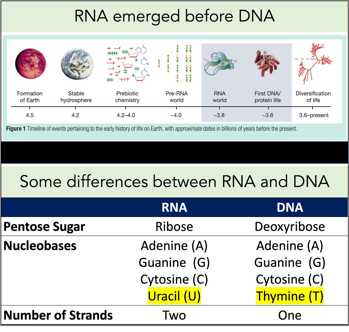
RNA came first. Then, ~3-4 billion years ago, DNA emerged.
Among their differences:
🔹RNA contains uracil
🔹DNA contains thymine
But why does DNA contains thymine (T) instead of uracil (U)?
https://t.co/XlxT6cLLXg
3/
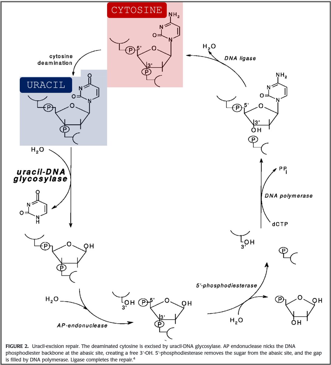
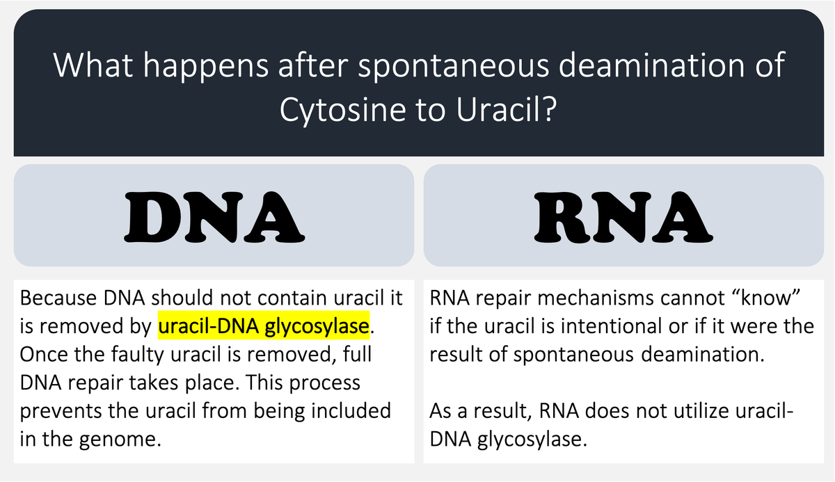
🔑Cytosine (C) can undergo spontaneous deamination to uracil (U).
In the RNA world, this meant that U could appear intensionally or unintentionally. This is clearly problematic. How can you repair RNA when you can't tell if something is an error?
https://t.co/bIZGviHBUc
4/
DNA's use of T instead of U means that spontaneous C → U deamination can be corrected without worry that an intentional U is being removed.
DNA requires greater stability than RNA so the transition to a thymine-based structure was beneficial.
https://t.co/bIZGviHBUc
5/
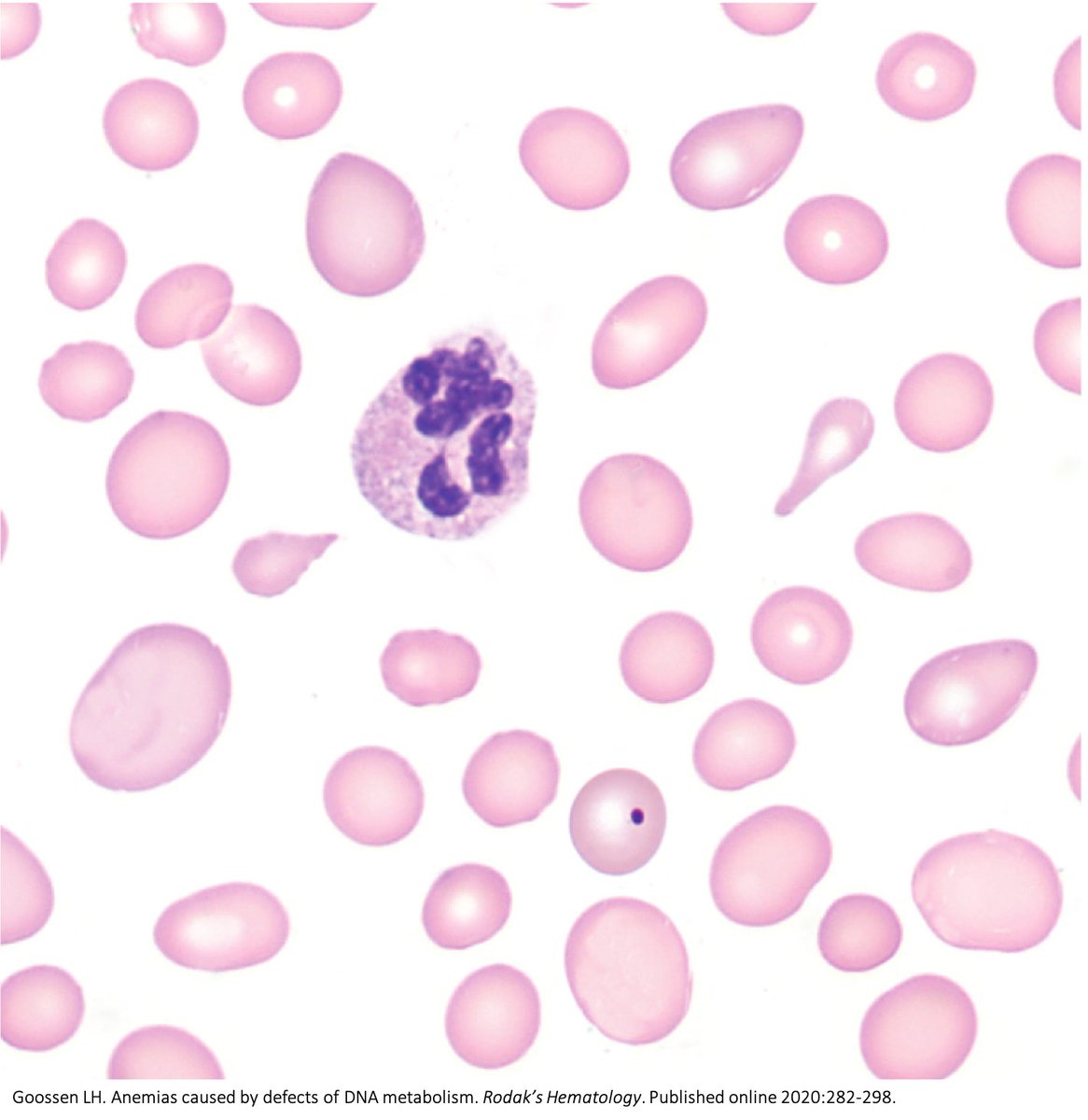
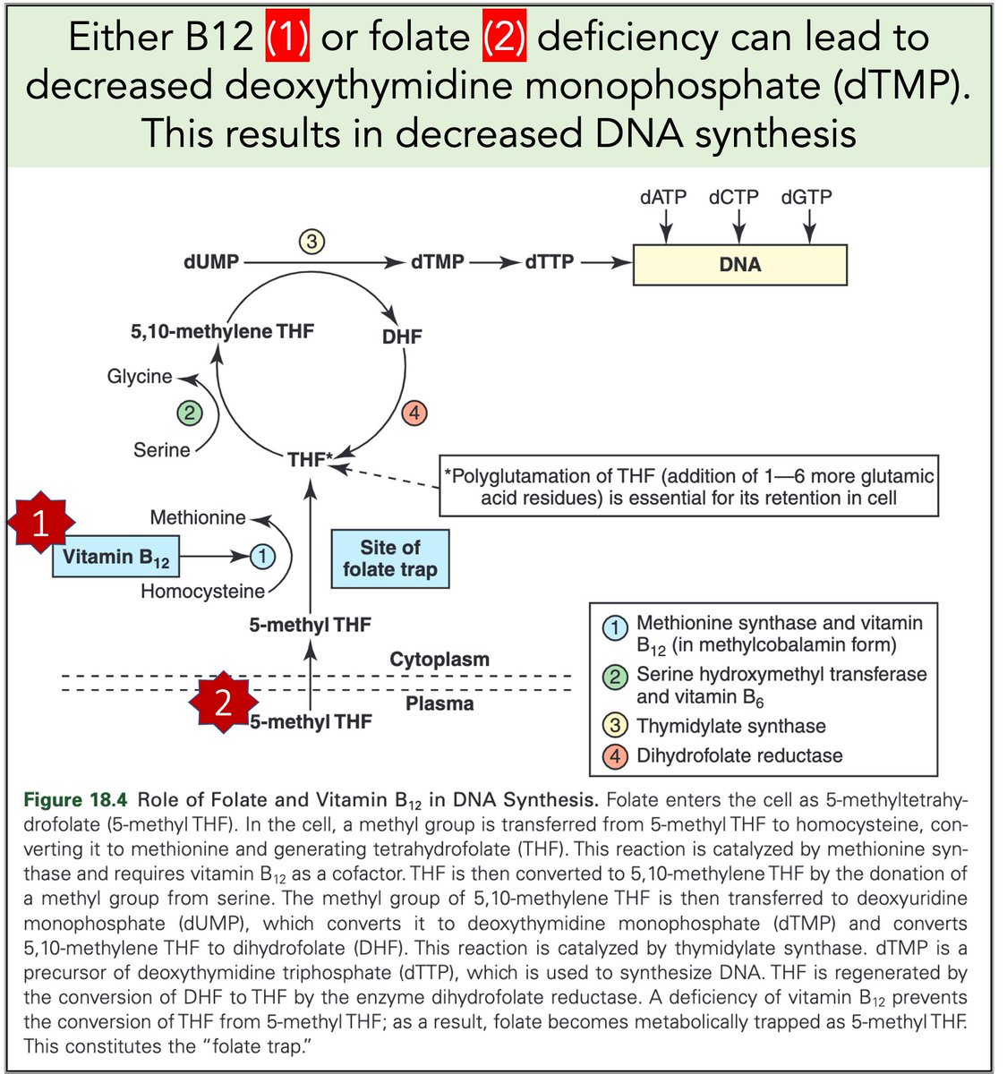
Let's return to megaloblastic anemia secondary to B12 or folate deficiency.
When either is severely deficient deoxythymidine monophosphate (dTMP*) production is hindered. With less dTMP, DNA synthesis is abnormal.
[*Note: thymine is the base in dTMP]
https://t.co/AnDUtKkbZh
Why do B12 and folate deficiencies lead to HUGE red blood cells?
And, if the issue is DNA synthesis, why are red blood cells (which don't have DNA) the key cell line affected?
For answers, we'll have to go back a few billion years.
2/
RNA came first. Then, ~3-4 billion years ago, DNA emerged.
Among their differences:
🔹RNA contains uracil
🔹DNA contains thymine
But why does DNA contains thymine (T) instead of uracil (U)?
https://t.co/XlxT6cLLXg
3/
🔑Cytosine (C) can undergo spontaneous deamination to uracil (U).
In the RNA world, this meant that U could appear intensionally or unintentionally. This is clearly problematic. How can you repair RNA when you can't tell if something is an error?
https://t.co/bIZGviHBUc
4/
DNA's use of T instead of U means that spontaneous C → U deamination can be corrected without worry that an intentional U is being removed.
DNA requires greater stability than RNA so the transition to a thymine-based structure was beneficial.
https://t.co/bIZGviHBUc
5/
Let's return to megaloblastic anemia secondary to B12 or folate deficiency.
When either is severely deficient deoxythymidine monophosphate (dTMP*) production is hindered. With less dTMP, DNA synthesis is abnormal.
[*Note: thymine is the base in dTMP]
https://t.co/AnDUtKkbZh

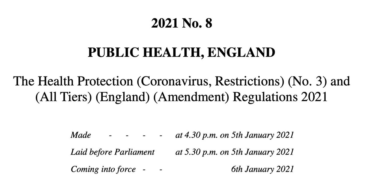
🚨New lockdown regulations just published, in force tomorrow
The Health Protection (Coronavirus, Restrictions) (No. 3) and (All Tiers) (England) (Amendment) Regulations 2021
https://t.co/L5jwlTDaIE
(Thread)
These are not a new set of regulations: they are amendments an old set of regulations
Which we thought were gone! But they are back
Welcome back No.3 regulations
A quick thing before we continue!
I have been analysing these laws for free for 9 months now - if you want to say thanks and have a few £ to spare please give to my @LawCentres fundraiser
They give free legal advice to people who need it
They also amend the All Tiers regulations
Oh god it's all amendments by paragraph references
Basically all of England now in Tier 4 and Tier 4 is amended but not by a huge amount
This really is a terrible way to make laws on the fly - who can possibly understand it?!
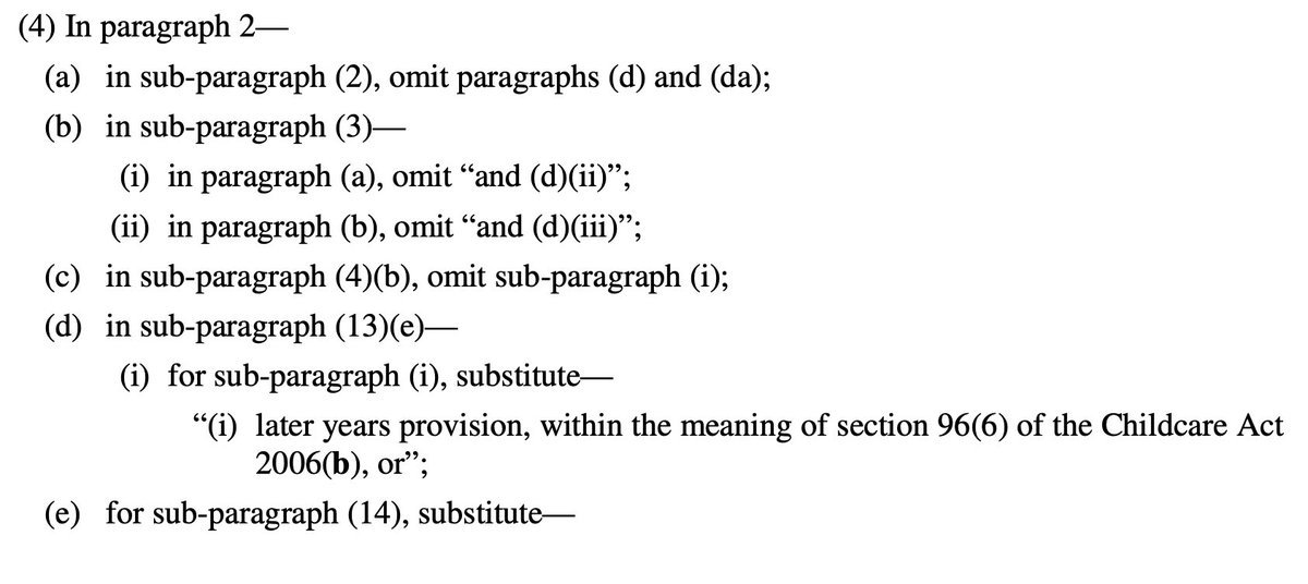
So, to explain, you need 2 documents open if you want to understand what is going on:
All Tiers regulations (Tiers 1-4, 2 December as amended) https://t.co/IraPQ112ak
And amendments https://t.co/L5jwlTDaIE
No sensible way of doing except by track changes, on it now, back soon
The Health Protection (Coronavirus, Restrictions) (No. 3) and (All Tiers) (England) (Amendment) Regulations 2021
https://t.co/L5jwlTDaIE
(Thread)
These are not a new set of regulations: they are amendments an old set of regulations
Which we thought were gone! But they are back
Welcome back No.3 regulations
A quick thing before we continue!
I have been analysing these laws for free for 9 months now - if you want to say thanks and have a few £ to spare please give to my @LawCentres fundraiser
They give free legal advice to people who need it
They also amend the All Tiers regulations
Oh god it's all amendments by paragraph references
Basically all of England now in Tier 4 and Tier 4 is amended but not by a huge amount
This really is a terrible way to make laws on the fly - who can possibly understand it?!
So, to explain, you need 2 documents open if you want to understand what is going on:
All Tiers regulations (Tiers 1-4, 2 December as amended) https://t.co/IraPQ112ak
And amendments https://t.co/L5jwlTDaIE
No sensible way of doing except by track changes, on it now, back soon


Some thoughts on this: Firstly, it might be personal preference, but I am not keen on this kind of campaign as I feel like it trivialises cancer. Sometimes the serious message gets lost because people are sharing pics of cats or whatever and the important context is gone.
More importantly, the statistic being used in the campaign is misleading. It says 57% of women put off cervical screening if they can't get waxed. But on further investigation, that's not accurate.
The page here goes on to say "57% of women who regularly have their pubic hair professionally removed would put off attending their cervical screening appointment if they hadn’t been able to visit a beauty salon."
So the 57% represents a concern not across the whole population of women, but only those who regularly get waxed. So how big of an issue is this across the whole population? And what else is stopping people getting smears?
I think campaigns for cancer screening are really tricky because there is so much nuance that often doesn't fit into a catchy headline or hashtag. It's certainly not easy and is part of a bigger conversation.
It\u2019s #CervicalCancerPreventionWeek \U0001f499
— myGP (@myGPapp) January 18, 2021
Here\u2019s how you can help to raise awareness:
\U0001f431 Share an image of the cat that best reflects your undercarriage/flower/bits (technical term vulva!) current look.
#\u20e3Use the Hashtag #myCat.
\U0001f46dTell and tag your friends to let them know. pic.twitter.com/8aHf96ynjT
More importantly, the statistic being used in the campaign is misleading. It says 57% of women put off cervical screening if they can't get waxed. But on further investigation, that's not accurate.
The page here goes on to say "57% of women who regularly have their pubic hair professionally removed would put off attending their cervical screening appointment if they hadn’t been able to visit a beauty salon."
So the 57% represents a concern not across the whole population of women, but only those who regularly get waxed. So how big of an issue is this across the whole population? And what else is stopping people getting smears?
I think campaigns for cancer screening are really tricky because there is so much nuance that often doesn't fit into a catchy headline or hashtag. It's certainly not easy and is part of a bigger conversation.