Two of those are linked to separate outbreaks of their own (a different LTC and a mental health hospital). Testing underway.
ICYMI, public health officials fear community transmission of B.1.1.7 (the "UK variant") is underway in Ontario.
I'll dissect each piece of worrying news from this story.
Two of those are linked to separate outbreaks of their own (a different LTC and a mental health hospital). Testing underway.
42 household contacts of ppl linked to the outbreak are also COVID+. Public health unit is worried about keeping cases (which are all likely B.1.1.7) from rippling further into the community.
https://t.co/81h7PtyNx2
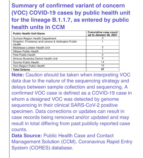
Here's a breakdown by health unit courtesy PHO:
https://t.co/87Czw5uYRD )
“If it isn’t spreading readily in our community now, it may very well do so in the near future."
@BogochIsaac: "The horse is out of the barn."
So what do we do now?
Bogoch: “There’s no magic, right? We know how to control this."
https://t.co/0qSObJike5
Dr. Gardner also spoke about restricting travel *inside* the province, between communities -- which he says would be controversial but worked in Australia.
https://t.co/0eViOe7ifT
Stay tuned.
- Now 85 secondary contacts from Roberta Place (up from the 42). h/t @LapointeShaw
- Nine of York Region's 15 B.1.1.7 cases acquired in community:
https://t.co/tZWTewVFaR
More from Health


Sarcomeres in cardiac myocytes (heart muscle cells) are mechanically coupled to focal adhesions through dorsal stress fiber-like structures. #cardiotwitter #CellBiology
1/13
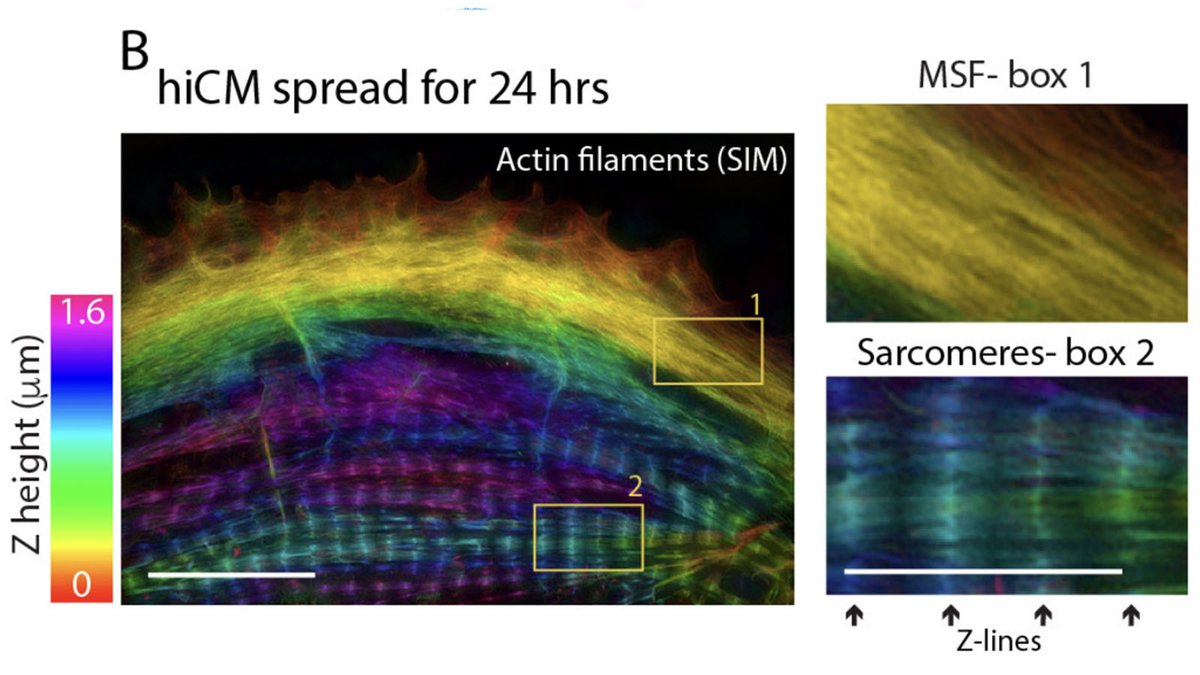
A thread based on Figure 1
A mature adult cardiac myocyte is packed with sarcomeres, whose contractile forces are coupled to the extracellular environment. With sarcomeres so close to the plasma membrane, how can we study the nature of this coupling?
2/13
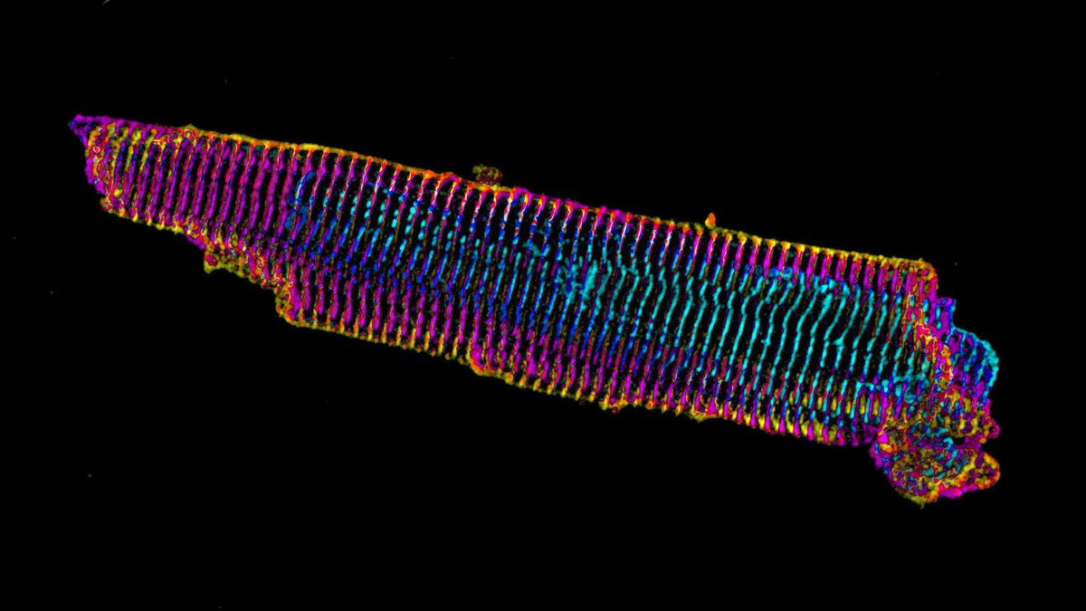
Short answer: find a model system where the sarcomeres are not so close to what the cardiac myocyte is attached to. Enter, iPS cell-derived cardiac myocytes. These are “immature” in culture as they resemble fetal or neonatal cardiac myocytes.
3/13
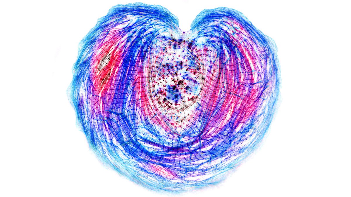
Our previous work on iPS cardiac myocytes reported that sarcomere containing myofibrils assembled on the top surface of the myocyte.
https://t.co/xIBCu3hG1W
4/13
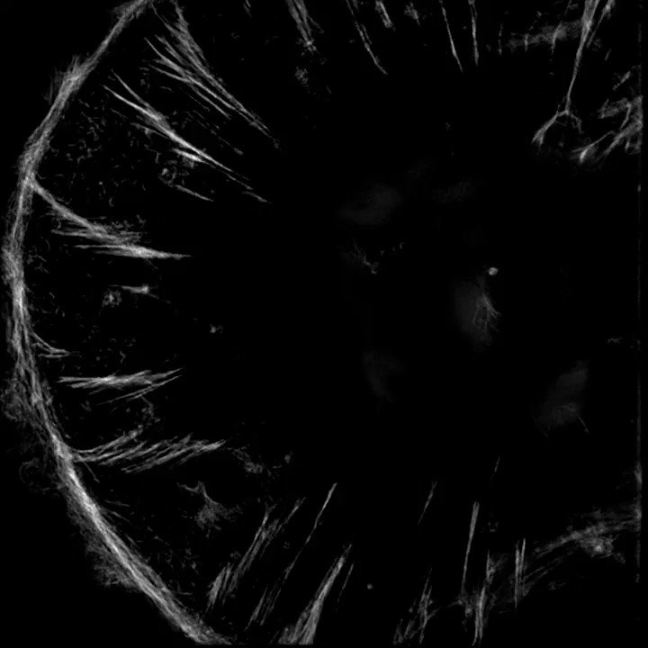
The sarcomeres seemed to be connected to focal adhesions on the bottom of the cell by thin actin bundles that resembled the dorsal stress fibers (DSF) commonly found in non-muscle cells. This movie steps through a Z stack of a myocyte starting at the bottom of the cell.
5/13
1/13
A thread based on Figure 1
A mature adult cardiac myocyte is packed with sarcomeres, whose contractile forces are coupled to the extracellular environment. With sarcomeres so close to the plasma membrane, how can we study the nature of this coupling?
2/13
Short answer: find a model system where the sarcomeres are not so close to what the cardiac myocyte is attached to. Enter, iPS cell-derived cardiac myocytes. These are “immature” in culture as they resemble fetal or neonatal cardiac myocytes.
3/13
Our previous work on iPS cardiac myocytes reported that sarcomere containing myofibrils assembled on the top surface of the myocyte.
https://t.co/xIBCu3hG1W
4/13
The sarcomeres seemed to be connected to focal adhesions on the bottom of the cell by thin actin bundles that resembled the dorsal stress fibers (DSF) commonly found in non-muscle cells. This movie steps through a Z stack of a myocyte starting at the bottom of the cell.
5/13